The Oxygen-Rich Postnatal Environment Induces Cardiomyocyte Cell-Cycle Arrest through DNA Damage Response
Feb 18, 2025
Highlights
- Mitochondrial mass and activity significantly increase in the heart after birth.
- ROS and DNA damage response concomitantly increase in cardiomyocytes postnatally.
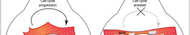
- Hypoxia delays cardiomyocyte cell-cycle arrest, whereas hyperoxia potentiates it.
- Scavenging ROS or inhibiting DNA damage response delays cell-cycle arrest
Summary
The mammalian heart has a remarkable regenerative capacity for a short period of time after birth, after which the majority of cardiomyocytes permanently exit cell cycle. We sought to determine the primary postnatal event that results in cardiomyocyte cell-cycle arrest. We hypothesized that transition to the oxygen-rich postnatal environment is the upstream signal that results in cell-cycle arrest of cardiomyocytes. Here, we show that reactive oxygen species (ROS), oxidative DNA damage, and DNA damage response (DDR) markers significantly increase in the heart during the first postnatal week. Intriguingly, postnatal hypoxemia, ROS scavenging, or inhibition of DDR all prolong the postnatal proliferative window of cardiomyocytes, whereas hyperoxemia and ROS generators shorten it. These findings uncover a protective mechanism that mediates cardiomyocyte cell-cycle arrest in exchange for utilization of oxygen-dependent aerobic metabolism. Reduction of mitochondrial-dependent oxidative stress should be an important component of cardiomyocyte proliferation-based therapeutic approaches.
Introduction
The pathophysiological basis of heart failure is the inability of the adult heart to regenerate lost or damaged myocardium, and although limited myocyte turnover does occur in the adult heart, it is insufficient for restoration of contractile dysfunction (Bergmann et al., 2009; Hsieh et al., 2007; Laflamme et al., 2002; Nadal-Ginard, 2001; Quaini et al., 2002). In contrast, the neonatal mammalian heart is capable of substantial regeneration following injury through cardiomyocyte proliferation (Porrello et al., 2011b, 2013), not unlike urodele amphibians (Becker et al., 1974; Flink, 2002; Oberpriller and Oberpriller, 1974) or teleost fish (González-Rosa et al., 2011; Poss et al., 2002; Wang et al., 2011). However, this regenerative capacity is lost by postnatal day 7 (Porrello et al., 2011b, 2013), which coincides with cardiomyocyte binucleation and cell-cycle arrest (Soonpaa et al., 1996). Although several regulators of cardiomyocytes cell cycle postnatally have been identified (Bersell et al., 2009; Chen et al., 2013; Eulalio et al., 2012; Mahmoud et al., 2013; Porrello et al., 2011a; Sdek et al., 2011; Xin et al., 2013), the upstream signal that causes permanent cell-cycle arrest of most cardiomyocytes remains unknown.
One of many factors shared by organisms that are capable of heart regeneration is the oxygenation state. For example, the zebrafish’s stagnant and warm aquatic environment has 1/30th oxygen capacitance compared to air and is prone to poor oxygenation, which may explain the remarkable tolerance of zebrafish to hypoxia (Rees et al., 2001; Roesner et al., 2006). Typical air-saturated water has a PaO2 of 146 mm Hg and zebrafish can tolerate hypoxia at PaO2 of 15 mm Hg (10% air-saturation) for 48 hr and even 8 mm Hg with hypoxic preconditioning. Moreover, the zebrafish circulatory system is relatively hypoxemic, as it has a primitive two-chamber heart with one atrium and one ventricle, which results in mixing of arterial and venous blood.
Cont. Reading Here